|
What is the OCULUS Keratograph?
The OCULUS Keratograph® 5M is an advanced corneal topographer with a built-in real keratometer and a color camera optimized for external imaging. Unique features include examining the meibomian glands, non-invasive tear film break-up time and the tear meniscus height measurement and evaluating the lipid layer. |

|
Keratograph Crystal Tear Report
The Keratograph helps determine the cause of dry eye disease quickly and reliably with the assistance of the new Crystal TEAR Report in the Keratograph® 5M. The Crystal TEAR Report includes 4 comprehensive scans:
The Keratograph helps determine the cause of dry eye disease quickly and reliably with the assistance of the new Crystal TEAR Report in the Keratograph® 5M. The Crystal TEAR Report includes 4 comprehensive scans:
- Tear Meniscus Height
- NIKBUT (non-invasive tear film break-up time)
- Redness Scan (R-Scan)
- Meibomian (oil glands) Eye Lid Scan

Meniscus Tear Height Assessment of the Tear Film Quantity
The height of the tear meniscus can be precisely measured with an integrated ruler and various magnification options, and its development along the edge of the bottom lid can be assessed.
Meniscus Tear Height Assessment of the Tear Film Quantity
The height of the tear meniscus can be precisely measured with an integrated ruler and various magnification options, and its development along the edge of the bottom lid can be assessed.
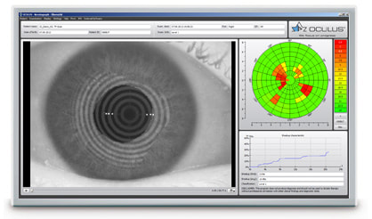
NIKBUT Assessment of the Tear Film Break-Up Time
The tear film break-up time is measured non-invasively and fully automatically. This scan is done by recording the way tears break up over the course of a few seconds without blinking. The infrared illumination is not visible to the human eye and prevents glare during the examination.

R-Scan Automatic Classification of Redness
Automatic classification of the bulbar redness. The R-Scan is the first module that automatically and objectively documents and classifies the bulbar and limbal degree of redness. The R-Scan detects the blood vessels in the conjunctiva and evaluates the degree of redness.